Novel optical methods for the investigation of living objects
We develop new optical methods for various life science applications, focusing on fundamental cellular processes.
These methods include:
• the development of new microscopy systems such as electro-optical microscopy,
• the use of original fluorescence microscopes to follow the dynamics of intra-neuronal transport (lien vers page DMD), or eukaryotic translation (lien vers page traduction),
• a new strategy for moving nanoparticles with light,
• the development and characterization of new optically active nanoprobes, some coupled with plasmonic nanostructures to improve their response.
Our activities are also linked to clinical research by
• the development of new methods in optometry, in close collaboration with industrialists in the field.
Eukaryotic translation kinetics
Permanent staff : Karen Perronet, François Marquier
We study the mechanisms of protein translation unsing a single-molecule fluorescence microscope that follows single ribosome kinetics. We can quantify initiation and elongation speeds of eukaryotic ribosomes.
High temporal and spatial resolution for nanoparticles tracking.
Permanent staff: François Marquier, Karen Perronet
Our objective is to develop a microscopy device that can address the measurement of intraneuronal transport in 3D, with a spatial resolution of less than 20 nm, and at temporal resolutions of less than a millisecond.
Novel optically active nanoprobes and enhanced optical properties
Permanent Staff: Gaëlle Allard, François Marquier, Karen Perronet, François Treussart
Our group is interested in the development of new types of probes for the measurement of biological processes at the molecular and nanometric scales. Part of this activity is also aimed at improving the optical properties of these particles to make them more efficient. Strong collaborations with laboratories in the Paris region give us access to many fabrication and characterization facilities.
New methods in optometry and vision science
Permanent staff : Richard Legras
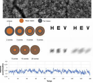
Optometry and Vision Science group is interested in physiological optics. One of the topics is the study of the quality of the retinal image in an attempt to understand more completely the limitations imposed by optical constraints on visual performance under various conditions. We used an adaptive optic system to modulate (correction and/or introduction) the eye’s monochromatic aberrations and measure visual performances in these conditions of aberrations. The numerical simulation of retinal image and visual performances are of particular interest. We are also centred in the study of the effect of novel methods for the correction of refractive error by spectacles, contact lenses, intra-ocular lenses or refractive surgery. We also work on the predictions of the subjective depth-of-focus and the through-focus visual acuity and contrast sensitivity curves, and on the adaptation to monochromatic aberrations.
Retinal imaging is also a subject of interest. We use a flood-illuminated AO retinal camera to image the retina. We constructed a large database on a healthy population. We also investigated the effects on axial elongation on the retinal structure.
More recently, the study of the fluctuations of accommodation is on particular interest.
Members
Marie-charlotte Chandeclerc

Marie-charlotte Chandeclerc
Doctorante sur "Real-time tracking of single nanoparticles motion and rotation in neurons"


